

3 min read
Osteoarthritis (OA) is a widespread condition that affects greater than 70% of the elderly population and poses a heavy cost burden on healthcare. It is a chronic degenerative disease characterized by the loss of articular cartilage components, which affects the entire joint structure. One of the major complaints by OA patients is the loss of joint function as well as chronic pain. Current therapies are focused on alleviating joint pain, however full pain relief is rarely experienced and significant side affects are commonly present. Research is not only focused disease pathology but also on understanding the mechanisms responsible for induction and maintenance of pain states.
The monosodium iodoacetate (MIA) model has been well characterized for the evaluation of OA pain. The iodacetate creates local inflammation in the knee from fluid expansion of the synovial mebrane, which is then followed by the production of inflammatory mediators (TNF-a, IL-1b, IL-6 and NGF). These mediators further contribute to OA pathogenesis by increasing cartilage degradation. The cartilage damage and degradation leads to chronic neuronal damage with neuropathic characteristics. The MIA-OA model then is thought to contain both inflammatory and neuropathic pain-related states by using local tissues and sensory innervation of the peripheral nervous system.
Inflammatory-related Pain State
CGRP (calcitonin gene-related peptide) is elevated in the sensory neurons innervating the knee and DRG (dorsal root ganglia). CGRP is an inflammatory driven neuronal marker whose expression is dependant on NGF (nerve growth factor) expression. The elevation of CGRP produces hyperalgesia and a pain sensation derived from inflammation.
Neuropathic-related Pain State
It is also suggested that proliferation of microglia in the dorsal horn of the spinal cord along with elevated levels of pro-inflammatory cytokines contributes to neuronal damage. The neuronal damage gradually rises along with the progress cartilage damage and this may lead to the chronic neuropathic pain state.
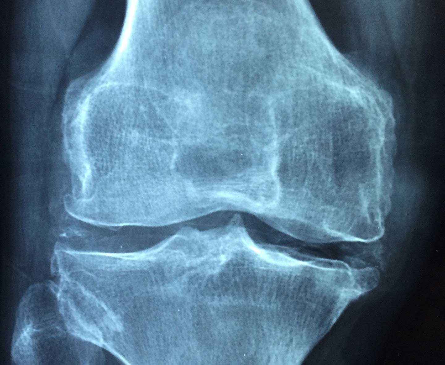
MIA-induced OA model
The MIA-induced OA model can allow the evaluation of anti-inflammatories (to treat early symptoms) as well as analgesics (to treat chronic pain). The study can be run anywhere from 14 days out to 56 days or longer depending on the proposed therapy.
Outcome measures and Readouts:
- Weight bearing measurements: weight bearing measures the spontaneous pain as a result of nerve injury or inflammation.
- Open field: the open field test would measure the animals desire to walk, the distance and speed if they did walk.
- Rotorod: the rotorod test would measure the ability to withstand the pain in a situation where walking was required.
- Joint compression: Randall Selitto can be used to measure the pressure required to withdraw (compression threshold)
- Histology and xray analysis of the knee
- Biomarkers (mRNA and protein level)
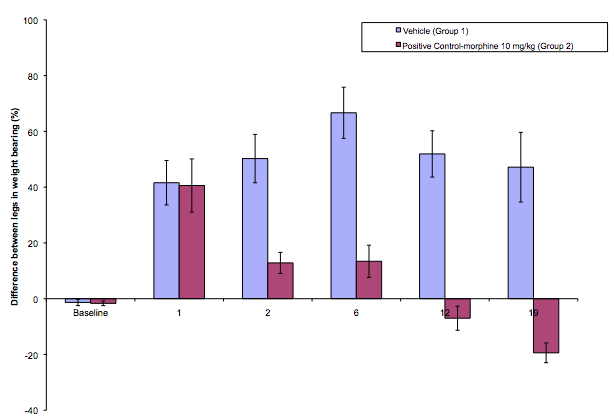
Weight bearing (% difference between left and right leg) of gapabentin and vehicle control in the MIA-induced model of OA.
- Orita, et al. (2011) Pain-related sensory inervation in monoiodoacetate-induced osteoarthritis in rat knees that gradually develops neuronal injury in addition to inflammatory pain. BMC Musculoskeletal Disorders 12:134
- Harvey, V and A. Dickenson (2009) Behavioral and electrophysiological characterisation of experiementally induced osteoarthritis and neuropathy in C57BL/6 mice. Molecular Pain 5:18
Related Posts
A minipig Model of Incisional pain
The use of animal models for the research of post-operative pain (POP) has been well described[1]....