Behavior
Conduct extensive pain, sensory, motor, and cognitive behavioral testing.


The figure shows grip test results in the MCAo stroke model, revealing significant differences between young and aged males, as well as between aged males and aged females.


Conduct extensive pain, sensory, motor, and cognitive behavioral testing.
Explore inflammatory and pain biomarkers in disease-specific tissues.
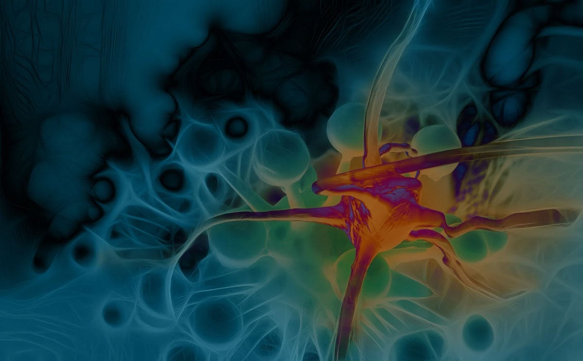
Characterize tissue and cellular changes in disease, pain, and neurodegeneration.