
Biomarker Analysis
Quantitative methods for translational markers in preclinical and clinical discovery.


Whitepaper: Translational Biomarkers
This whitepaper provides an overview of how biomarkers can be utilized to extract maximum value early in translational studies while being optimized for use in clinical studies.

Whitepaper: Inflammatory Biomarkers
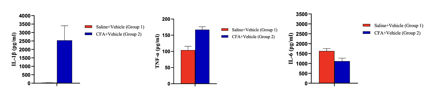
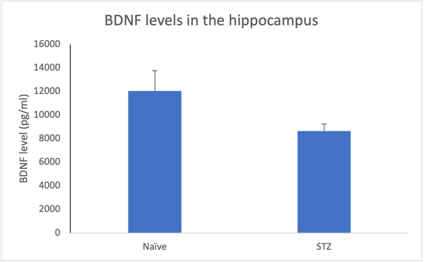
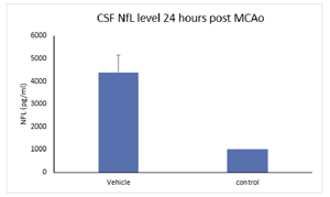
The interplay between the central and peripheral nervous systems and the immune response highlights the importance of understanding the cellular and inflammatory attributes that are strongly associated with neurological diseases and pain.

Whitepaper: High Precision Biomarker Detection
Multiplex and ultra-sensitive detection assays provide a powerful platform for difficult to measure analytes. The detection of these markers offers invaluable insight on monitoring disease progression at earlier stages and assessing the need for therapeutic intervention.
