
Behavior
Conduct extensive pain, sensory, motor, and cognitive behavioral testing.


Conduct extensive pain, sensory, motor, and cognitive behavioral testing.
Explore inflammatory and pain biomarkers in disease-specific tissues.
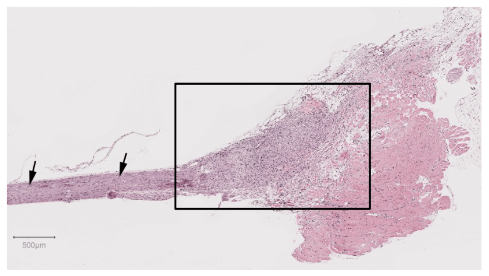
Characterize tissue and cellular changes in disease, pain, and neurodegeneration.
Measure motor and sensory evoked potentials to assess disease progression and pain.


