

2 min read
Experimental autoimmune encephalomyelitis (EAE) is one of the most widely used and well-characterized models for studying multiple sclerosis (MS). Although no animal model fully replicates the complexity of the human disease, EAE has been instrumental in advancing the understanding of immune-mediated neuroinflammation, demyelination, and neurodegeneration. More than half of the MS drugs currently approved were originally studied in EAE models, underscoring its value as a preclinical platform.
EAE can be induced using different antigens such as myelin oligodendrocyte glycoprotein (MOG), proteolipid protein (PLP), or myelin basic protein (MBP). Each of these models mimics distinct facets of MS pathology, making them adaptable tools for exploring immune responses, neuroprotective strategies, and therapeutic efficacy. A range of assessments is available across these models, including clinical scoring of motor function, biomarker profiling through cytokine analysis and pharmacokinetics, histological and immunohistochemical examination of demyelination, and behavioral measurements that capture motor, sensory, pain, and cognitive changes.
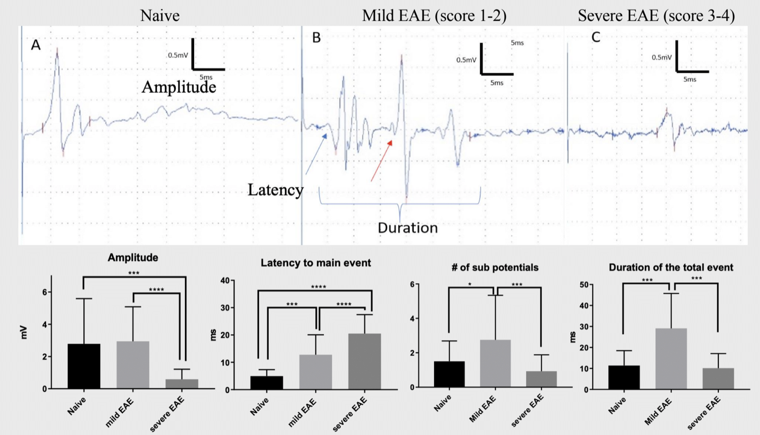
Traditionally, clinical scoring has been the primary means of evaluating disease progression in EAE. However, this method has limitations. Clinical scores often plateau after peak disease, making it difficult to differentiate between treatment groups or to capture subtler aspects of disease progression. To address this gap, electrophysiological recordings have emerged as a powerful complementary approach. Motor evoked potentials (tcMEPs) provide objective data that distinguish between mild and severe disease states, while sensory evoked potentials (tcSEPs) reveal early functional changes that are not detected by clinical scores alone.
The P25 Wave: An Early Sensory Biomarker
One of the most intriguing electrophysiological findings in MOG-induced EAE is the identification of the P25 sensory wave. This novel biomarker appears before clinical signs develop, around day 10 post-induction, and correlates with pain-related behavior. The P25 amplitude increases during the early stages of disease, then declines as severity progresses, even while clinical scores remain constant. Importantly, elevated P25 has been observed in some animals that never developed observable clinical symptoms, suggesting that subclinical pathology may be present. This mirrors what is suspected in MS patients, where disease activity is thought to begin years before diagnosis.
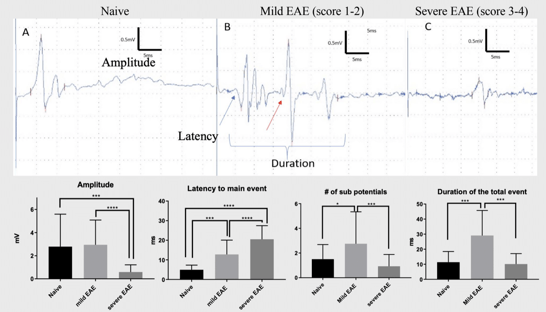
The discovery of P25 highlights the importance of integrating electrophysiology into EAE studies. By providing sensitive, objective measures of both motor and sensory pathway function, electrophysiology enables earlier detection of disease, better differentiation of severity, and more comprehensive evaluation of treatment effects. Combined with biomarkers, histology, and behavioral testing, this approach expands the utility of EAE models beyond clinical scoring alone, offering a more nuanced view of disease mechanisms and therapeutic response.
View the full publication here.
Reference:
Sefiani A, et al. A Novel Sensory Wave (P25) in Myelin Oligodendrocyte Glycoprotein-induced Experimental Autoimmune Encephalomyelitis Murine Model. Journal of Pain.
Related Posts
Glatiramer Acetate (Copaxone® Generics) and Batch Release Testing in MS/EAE at MD Biosciences
MDB Gains OECD-GLP certification for In Vivo Glatiramer Acetate Batch Release Testing Services
Monitoring pain in EAE animals experiencing paralysis
Many neurodegenerative diseases such as Multiple Sclerosis, ALS, and Rhett Syndrome involve...
Do clinical scores tell us all we need to know?
Multiple sclerosis is a disease in which the immune system attacks the myelin sheath that covers...