

4 min read

The goal of the preclinical development process is to help predict clinical efficacy, emphasizing the importance of studying diseases through models that yield predictive and clinically relevant data. This involves using both in vivo and in vitro approaches to understand the complexities of human diseases. In vitro models provide valuable insights into the mechanisms of action, while in vivo models offer translational outcomes.
In Vitro Modeling to Advance Neurodegeneration Research & Development
There are many advantages to cell-based assays, which are now gaining regulatory acceptance as part of the preclinical package for drug development. Cell-based assays are cost and time-effective and provide an advantageous model for high-throughput screening, safety, and efficacy. In vitro screening assays are ideal for evaluating compounds before testing in vivo efficacy. The results are highly reproducible and relevant to the clinic, especially when derived from human cells. In vitro models offer valuable insights into the mode of action of diseases, like Alzheimer's Disease, which are challenging to model in animals.
Neurite Outgrowth Assays
In vitro screening assays utilize primary neurons from immature mice or rats. These assays can then be conditioned and treated with compounds to evaluate endpoints for neurodegeneration. A hallmark of degeneration is the neurite blebbing, indicating neurite disassembly and degeneration. The neurodegeneration index (NI) compares the relative area of neurites and the number of blebs. Neurite outgrowth assays also allow for the examination of neural integrity, development, and the regenerative potential of neurons, which could be particularly relevant in studying spinal cord injury and the effect of psychedelic drugs on neurons.
Spinal cord injury can be replicated in adult tissue cultures in in vitro assays that mimics the human condition of SCI.
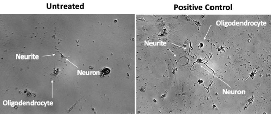
Neurite outgrowth. Untreated: small neurons, not much has developed with neurites and oligodendrocytes (left). Positive control with more developed neurons in dish with synapses created, long neurites.
Neurodegeneration Screening Assays
The neurodegeneration assay is an in vitro method to examine the potential effects of exogenous compounds on neuronal integrity. This assay provides valuable information on neuroprotection and can be used to screen multiple compounds at once. The neurodegeneration screening assay can be performed for diseases such as Alzheimer's, Parkinson's, diabetes, and chemotherapy-induced peripheral neuropathy (CIPN). Alzheimer's disease, for example, involves widespread changes, including the formation of plaques and tangles, synaptic dysfunction, and inflammation. Replicating these complex interactions in animals is more challenging than studying isolated cells in vitro. In this model, cortical neurons from mice are conditioned with beta-amyloid. This method can be replicated for a number of indications. For Parkinson's, cortical neurons are conditioned with alpha-synuclein.
Below is an example of an in vitro Parkinson's disease model: Cortical neurons from immature mice were conditioned for 7 days with a-synuclein. Cultures were treated with compound and stained with Beta-tubulin III antibody staining. This shows a reduction in neurite area and an increase in neurite blebbing, which is indicative of neurodegeneration.
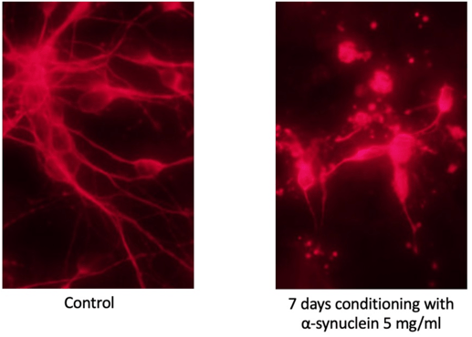
Parkinson's disease model. Learn more here.
Synaptic Imaging
Synaptic imaging assays evaluate the effects of treatments on synapse formation, synaptic strength, neurite length in early development, and synaptic density, all of which are important parameters for understanding neural networks. This assay advances cognition, learning, and degenerative disease research using primary hippocampal neurons obtained from mouse and rat models. The process involves imaging techniques that focus on the Synaptobrevin2 protein along with other synaptic markers.
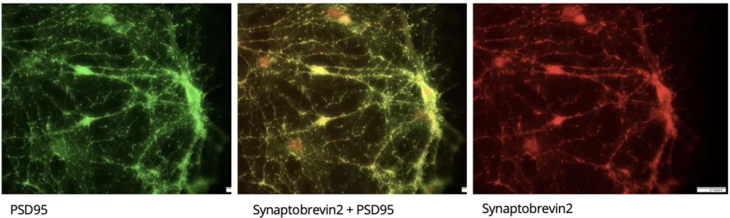
Synaptic imaging markers: PSD95 is a marker for the dendrite side of the synapse. Synaptobrevin2 is a vesicle membrane attached protein that is a marker for synapse numbers and characteristics.
iPSC Reprogramming
Induced pluripotent stem cells (iPSCs) can be characterized and differentiated to provide a customizable tool for researching neural development, disease modeling, drug screening, and target validation. iPSCs are reprogrammed to differentiate into neurons and enable neuronal toxicity and safety tests by measuring injury markers and performing cell viability assays. Cardiomyocyte iPSC assays allow for studying neurodegenerative-induced cardiac diseases, such as cardiac hypertrophy and cardiac arrhythmia. These assays also enable cardiac toxicity and safety tests.
To learn more about our models or to speak with a scientist, click here.
Related Posts
International Congress on Neuropathic Pain 2025
We had the opportunity to present at NeuPSIG 2025 in Berlin, sharing our latest findings in...
Successfully Predict Neuropathic Pain Conditions
The rodent has historically been used as the dominant model for the study of pain mechanisms and...
Assessing Subjective Pain | Preclinical Pain Studies
Rodent models of pain such as nerve injury models are important to understand the mechanisms that...