


3 min read
Quantitative Histology: What Computational Image Analysis Adds to a Preclinical Package
By: MD Biosciences on Jul 28, 2026 9:00:01 AM
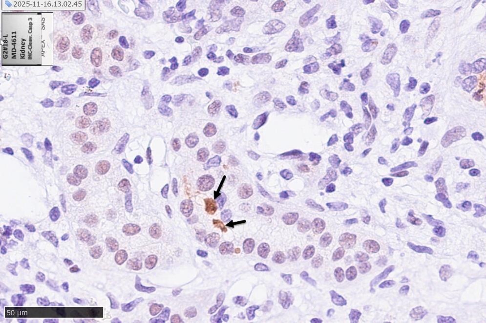
Histology has always carried a quiet tension. It is the most direct evidence a preclinical study can offer, a look at the actual tissue the candidate acted on, and at the same time it has historically been the least quantitative part of the package. A pathologist reading slides produces ordinal scores and expert judgment, both valuable, both subjective, and a graded scale of zero to four collapses a continuous biological reality into a handful of bins. For a confirmatory endpoint that a regulator or a development team will lean on, that subjectivity and that loss of resolution are real limitations. Computational image analysis is changing this, not by replacing the pathologist but by turning what the pathologist sees into continuous, reproducible, auditable measurements. The shift matters most when the endpoint is the basis for a decision rather than a descriptive footnote.
From Ordinal Scores to Continuous Measurements
The practical gain from a deep learning image analysis platform is that it reads entire sections consistently and returns numbers rather than categories. The same staining that a manual reader would grade can instead be quantified across the whole tissue, every structure of interest counted and measured the same way each time, which removes inter reader variability and reader fatigue from the dataset. Three applications illustrate the range. CD31 immunostaining marks blood vessels, and quantifying vessel density across a section turns angiogenesis from an impression into a measured value, which is central to wound healing and any program acting on vascular biology. Herovici staining differentiates collagen I from collagen III, and computational analysis of the ratio reports on the maturity and quality of new connective tissue rather than simply its presence, a distinction that matters when the question is whether a wound is healing well or merely closing. The throughput and consistency of automated reading also make it feasible to apply these measures at a scale that manual quantification would make impractical.
The Endpoint That Translates Directly: IENF Density
The clearest case for quantitative histology is intraepidermal nerve fiber density. In the clinic, IENF density measured from a skin punch biopsy is the reference standard for diagnosing small fiber neuropathy, which means a preclinical endpoint using the same measurement is reading the same quantity a clinician would. Measured with the pan-neuronal marker PGP9.5 and quantified by image analysis, IENF density becomes a translational bridge rather than a species specific proxy, and its sensitivity is real. In the Göttingen minipig postoperative pain model, IENF density was 40 percent higher in hind leg skin than in flank skin, 4.8 ± 1.3 against 3.4 ± 1.4 nerve fibers per mm², a difference that reached statistical significance (p<0.05) and that informs where a study should sample. An endpoint that can resolve a regional difference of that size in healthy tissue is an endpoint with the dynamic range to detect a treatment effect.
Multiple Antibodies, One Quantified Read of the Skin
The most developed expression of this approach is ChemoMorphometric Analysis, a proprietary method that quantifies multiple antibody markers across skin biopsies to produce an integrated profile rather than a series of isolated stains. Its translational credibility comes from how it was validated. In the porcine peripheral neuritis model, ChemoMorphometric Analysis of skin biopsies, reported by Rice and colleagues in 2019 in Neurobiology of Pain, found reduced intraepidermal nerve fiber density alongside increased CGRP, increased Nav1.7, increased endothelin A receptor, and decreased endothelin B receptor expression in keratinocytes. That constellation mirrors what is found in human neuropathic pain skin biopsies, and the concordance is detailed enough that it stands as one of the strongest pieces of translational validation in the porcine pain literature. The point is not that any single marker moved, but that a quantified, multi marker skin signature in the animal matched the human pathology marker by marker.
Why the Quantification Is Doing Real Work
It would be easy to read all of this as a workflow improvement, faster slides and prettier numbers, but the substantive value is in what continuous quantification enables that ordinal scoring cannot. Continuous measures have the statistical power to detect graded, dose dependent effects that a four point scale would flatten into no apparent difference. Reproducible, automated reads produce endpoints that can be audited and defended when a regulator or a partner asks how the number was derived. And the ability to quantify several markers across the same tissue lets a program build an integrated histological signature, the way ChemoMorphometric Analysis does for skin, rather than reporting disconnected stains. For wound healing, peripheral neuropathy, pain, and any program where the tissue level effect is the claim, that combination changes histology from supporting description into a primary, quantitative endpoint.
MD Biosciences runs quantitative histology on the DeePathology STUDIO platform, including CD31 vessel density, Herovici collagen typing, PGP9.5 intraepidermal nerve fiber density, and ChemoMorphometric Analysis of skin biopsies, integrated with the in vivo and biomarker work from the same animals. For sponsors considering whether a quantitative histological endpoint would strengthen a program, study design discussions are welcome at neuro@mdbiosciences.com.
References:
Rice FL et al. 2019. Human-like cutaneous neuropathologies associated with a porcine model of peripheral neuritis. Neurobiology of Pain. (Meilin co-author.)
Castel D, Schauder A, Aizenberg I, Meilin S. 2021. Validation of a Göttingen minipig model of postoperative incisional pain. Journal of Anesthesia and Surgical Care.
Related Posts
Three ways to approach translation
As the general population continues to age, the economic burden of chronic pain and...
Glatiramer Acetate (Copaxone® Generics) and Batch Release Testing in MS/EAE at MD Biosciences
MDB Gains OECD-GLP certification for In Vivo Glatiramer Acetate Batch Release Testing Services
Promoting Diversity for Translational Success
In preclinical efficacy studies, several factors are often overlooked, and it is important to...