

6 min read
As the general population continues to age, the economic burden of chronic pain and neurodegenerative diseases is increasing and becoming unsustainable. Unfortunately, the pipeline of new therapeutics has thinned over the past several years due to several clinical stage failures. Reasons for these failures may include insufficient understanding of neurobiological mechanisms, poor translation of preclinical data, clinical trial challenges, and the complexity and variability of the human brain and neurological diseases.
The intense medical need for new and better treatments for the aging population has led to neuroscience being one of the largest and fastest growing focus areas in drug development, predicted to take center stage in the coming years, a place currently occupied by oncology.
As drug developers race to develop the next therapeutic, many are looking to incorporate more translational approaches to their development strategy. In this article, we approach 3 ways drug developers can approach translation.
1. Using large animal species more often.
During preclinical stages, researchers utilize disease models that aim to replicate the human condition. In theory, these models would be able to replicate the human disease, share underlying biological mechanisms with human disease, and have predictive value in safety and efficacy in human.
These criteria are often unmet, or at least not met fully within one model.
Challenges often include:
- differences in basic biology between rodents and humans
- lack of homology with molecular targets and pathways
- lack of diversity in rodents used in research vs human
- comorbidities associated with age-related diseases in humans that are not reflected in rodent models
- environmental factors that contribute to human diseases not replicated in disease models.
Increasing the clinical translatability of preclinical data requires models that accurately reflect the human condition. Much of current research is conducted in rodents, but the anatomy and physiology that differs from humans. Rodents have lower brain function, shorter lifespan, smaller bodies, different immune mechanisms, less complex CNS systems etc.
While rodents provide valuable data and will always occupy a place in the process, drug developers must consider adding more non-rodent species that have greater similarities to the human condition when possible. Large animal models often reproduce key features of human biology and disease more closely. Non-human primates however are costly, difficult to access, and have long lead times. Due to this, attention has turned to pig models.
The pig shares valuable similarities with humans, including skin and neurological characteristics, nerve stimulation and behavioral changes. Consider some of the major challenges within pain research specifically to see how large animals can bridge the gap between preclinical and clinical studies.
- Sensory nerve function -TRPV1 is a non-specific cationic channel located on neuronal cells which innervate most of our organs. It acts as a multisensory receptor for potential injury signals and detects and integrates nociceptive and thermal stimuli in sensory nerve fibers. TRPV1 is highly involved in initiating inflammation and transmission of pain signals. The phylogenic tree of TRPV1 shows human and pig much more closely related than rodent.
- Skin innervation -The skin is a highly sensitive organ that is densely innervated with different types of sensory nerve endings. Intraepidermal nerve fiber analysis (IENF) shows a similar nerve density between human and pig whereas the rodent has a much higher density. We have also identified that there is no standard method for analyzing IENF in the rodents, whereas the analysis in the pig is done via similar methods as in the clinic. This adds to the difficulty in assessing the changes that are happening in the rodents.
- Skin structure and penetration properties - The hair density and epidermis-dermis ratio are much closer between pig and human than that of rodents. Healing in humans, as well as pigs, also occurs by re-epithelialization rather than contraction in the rodent. Additional similarities between pig and human is that there is an 80% predictability in permeability and greater than 93% similarity in biomarkers.
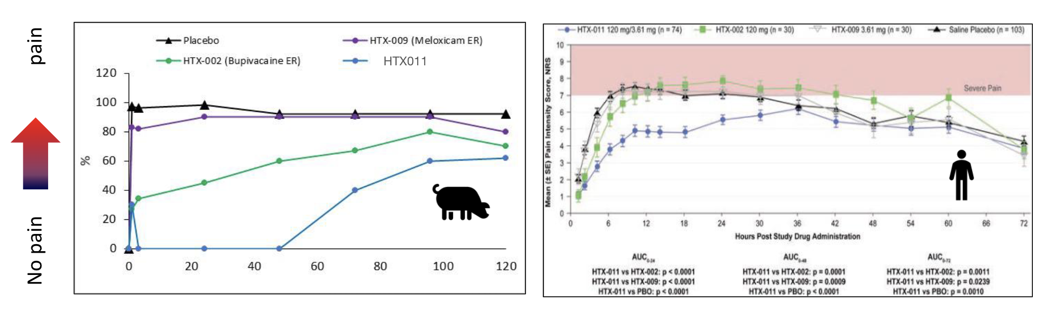
The data below shows an example of the predictability of the pig models. Meloxicam (HTX-009) and Bupivacaine (HTX-002) were evaluated in a flank incision model in the pig. Meloxicam shows no activity in the pig and almost no activity in the human as well. In contrast, Bupivacaine is slightly active in both the pig and human at the beginning. HTX-011 (a combination of Meloxicam and Bupivacaine) shows a synergistic effect in both pig and human.

2. Incorporating Translational Biomarkers
The use of biomarkers has the potential to increase value early in drug development by extracting more data and information from each study. This can increase efficiency, reduce costs, guide decisions, boost the translational power of preclinical studies, and effectively enable more drug candidates to move forward.
Between 2015-2019, over half of the approvals by the FDA (59%) and EMA (69%) were accompanied by at least one biomarker (source: clinicaltrials.gov). While advances have been made in biomarker development for other disorders (eg cancer), neuroscience biomarkers lag behind due to the complexity of the nervous system and access to tissue and the blood brain barrier.
A growing range of biological measures that include proteomic and cell-based signatures are being explored as potential biomarkers in the field of pain and neurodegenerative conditions. Drug developers can incorporate target biomarkers, mechanism biomarkers, and disease biomarkers into preclinical studies to objectively measure physiological processes, pathophysiological processes, or pharmacological responses.
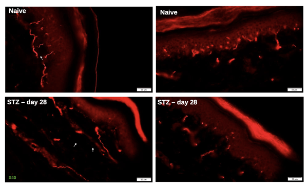
In the data below, Immunohistochemical analysis of intra-epidermal nerve fiber (IENF) density provides insight into changes in nerve fiber density between naive and STZ-diabetic animals. In the clinic, IENF density may serve as a histological biomarker of nociceptive nerve fiber involvement in peripheral neuropathy.
3. Incorporating cutting edge technologies used in humans.
Using outcomes that are comparable to those measured in patients, researchers can increase the translational value and predictability of preclinical studies.
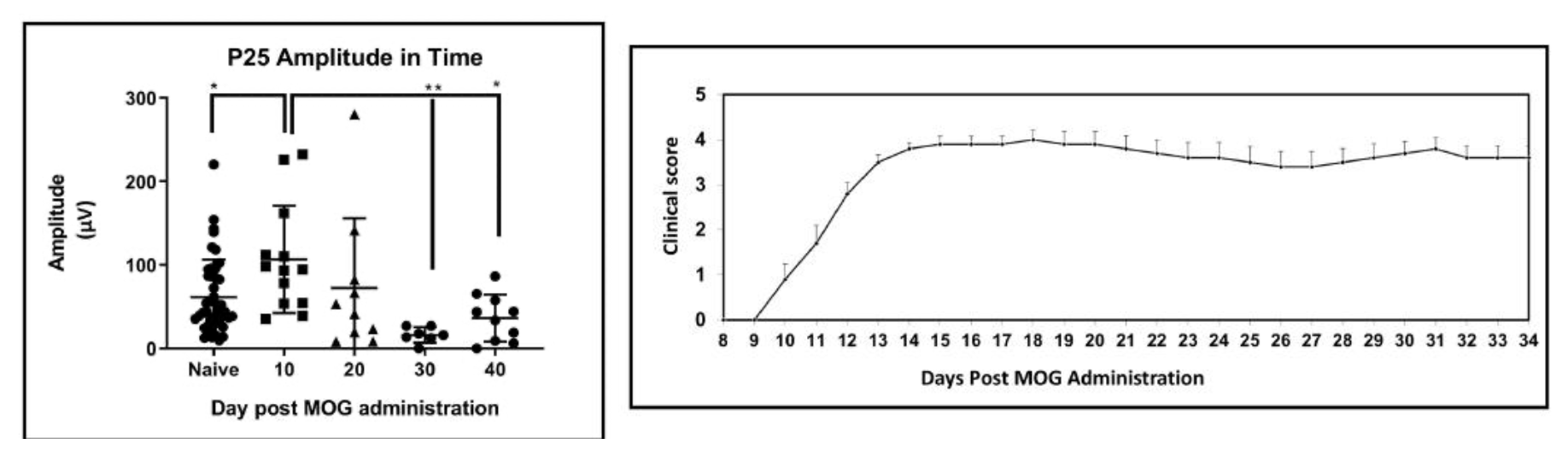
Electrophysiology (EP), an in vivo biomarker that is used in the clinic, provides the ability to study neurological dysfunctions, how the nervous system is affected, and how it responds to various treatments. Electrophysiology can be conducted to study the functional properties of neurons, which allows researchers to better understand the neurological disorder and the potential of neuroprotective or neurodegenerative therapies.
When adding electrophysiology to a preclinical study, researchers are able to see what is otherwise missed when relying on clinical scores alone. In the data example below, tcSEP (sensory evoked potentials) are monitored using electrophysiology. EP allows researchers to distinguish between mild and severe disease (using motor evoked potentials) and can be used as a diagnostic tool to identify early signs and symptoms before clinical scores appear.

As drug developers aim to improve the success rate of new CNS and pain therapies, the focus on translational approaches will increase. Just as with other indications, larger animal models, biomarker panels and clinical endpoints have the potential to transform neuroscience drug development and the medical landscape.
MD Biosciences, a preclinical neuroscience CRO, has the expertise to design programs that incorporate these translational approaches and help de-risk clinical studies.
Related Posts
The Critical Role of Clinical Relevance in Effective Study Design
Enhancing clinical relevance in early-stage studies can improve efficiency, reduce costs, and...
The HTX-011 Story
HTX-011 Breakthrough in Pain Management
ZYNRELEF, the commercial name for HTX-011, is the first and...
Advancing Nerve Injury and Repair Models
Understanding Mechanisms Underlying Neuropathic Pain