

2 min read
Spinal cord injury (SCI) remains one of the most challenging neurological conditions to treat, with few therapies successfully translating from the lab to the clinic. Studying SCI is challenging because it involves both acute mechanical injury and progressive neuroinflammation and degeneration. Selecting an appropriate model is critical to capture these different aspects. Yet many preclinical studies rely on a single model system, which contributes to translational failures.
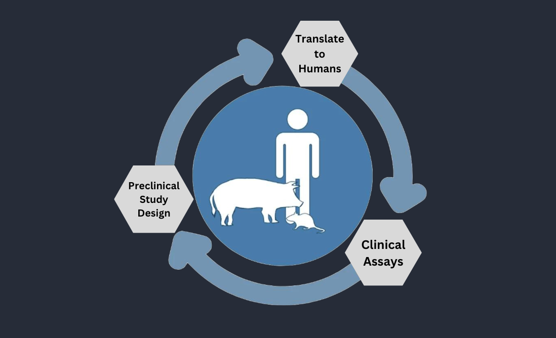
At MD Biosciences, we have developed a translational approach to SCI research that spans discovery to clinical translation. Our platform combines in vitro cell-based assays, rodent models, and advanced large-animal models.
Optimizing In Vitro Models
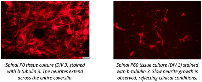
Traditional in vitro SCI models often use spinal cord tissue from young donors. While these cultures support rapid neurite outgrowth, they lack the primary components that inhibit neuronal growth in adults, making them less predictive of human outcomes. To better replicate clinical conditions, we use adult donor spinal cord tissue in our neurite outgrowth assays. Although neurite outgrowth takes longer in these cultures, the assay more accurately reflects the microenvironment of the glial scar. By measuring neurite density, length, and synapse formation, we gain a more reliable predictor of potential therapeutic success.
Rodent Models: Understanding Functional Recovery
Rodent models are widely used in SCI research. In preclinical studies of SCI, controlled contusion or transection injuries in mice are carefully evaluated over 12–24 weeks using behavioral scoring, gait analysis, and body weight tracking. Histopathology, cytokine analysis, and electrophysiology further inform the extent of functional loss and recovery. Electrophysiological assessments, such as motor evoked potentials (MEPs), allow us to quantify functional changes in spinal cord pathways after injury. Download the datasheet to learn more.
Porcine Models: Bridging the Gap to Human SCI
While rodent models provide critical mechanistic insights, pigs offer an unparalleled advantage for translational SCI research due to their anatomical and physiological similarities to humans. In our porcine models, SCI is induced via controlled contusion to replicate clinically relevant trauma. Recovery is monitored through advanced gait analysis, behavioral scoring, and electrophysiological assessments.
The pigs demonstrate recovery patterns that closely mimic clinical outcomes. Behavioral assessments capture changes in locomotor function over time, while body weight tracking serves as an overall indicator of health and recovery. Electrophysiology provides a powerful readout of spinal cord function, with partial injuries showing rapid MEP recovery and severe injuries reflecting significant functional loss.
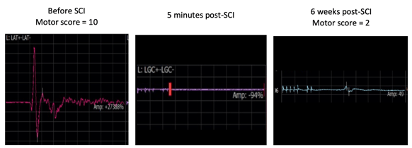
Figure 1. MEP recordings 5 minutes post-contusion show no detectable signal, indicating complete loss of function. By 6 weeks, minor signal recovery is observed, reflecting slight improvement consistent with expected outcomes in this model. Download the datasheet to learn more.
By combining in vitro adult tissue cultures, rodent models, and advanced porcine models, MD Biosciences is at the forefront of translational SCI research. Our integrated platform enables comprehensive evaluation of therapeutic interventions, providing models that help bridge the gap to clinical translation.
Contact us to learn more or receive a quote.
Related Posts
Sex Differences in Pain Research: What Your Preclinical Program May Be Missing
In 2016, the NIH mandate on sex as a biological variable (SABV) formalized what pain researchers...
New Developments: Innovative Cold Pain Testing Methods
Cold testing in rats serves as a crucial tool in pain research, particularly in studying...
Rethinking the Preclinical Package for Chronic Kidney Disease: Why Pigs belong in the conversation
Drug development for chronic kidney disease has entered a more ambitious phase. SGLT2 inhibitors,...