

5 min read

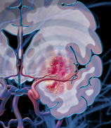 Post-stroke neuroinflammation is a very complex phenomenon involving multiple resident and invading cell types at varying degrees of differentiation or activation each expressing specific subsets of diffusible factors, receptors, cellular adhesion molecules, and other markers, all of which is changing as time passes to create an initially neurotoxic and then finally neuroprotective environment. This inflammatory process in the penumbra offers a broad array of potential cellular and molecular targets with much wider therapeutic windows. At the cellular level, neurons, microglia, astrocytes, and cerebrovascular endothelial cells are the first affected by the ischemic conditions and their responses to massive cell death in neighboring tissue initiates the precisely timed arrival of successive subsets of leukocytes – first neutrophils, followed by monocytes and macrophages, and finally T cells. Targeting these cells via manipulation of their phenotypes or activation states or their movements into lesion sites or their release of harmful mediators represents a major investigative pathway toward potential therapeutics for ischemic stroke sufferers. [1-4]
Microglia Microglia are the first to detect and respond to ischemic conditions, showing signs of activation as early as a few minutes after infarct. Microglia are the resident brain immunocompetent cells and constitutively express a wide variety of pattern recognition receptors. After stroke, resting microglia detect the spilled cytoplasmic contents of necrotic cells as well as high levels of ROS and consequently transform to an active, phagocytic phenotype, functioning to clear dead cells and cellular debris and release pro-inflammatory and cytotoxic diffusible factors. Inhibitors of microglial activation, such as the multi-functional drug Minocycline, have been shown to reduce infarct size in animal models when administered intraveneously in combination with rt-PA within 6 hours of infarct. At later timepoints, it appears microglia have neuroprotective effects, such as producing neurotrophic and other growth factors and supporting neurogenesis and neural plasticity. [1-4]
Astrocytes Astrocytes normally provide energy, neurotrophic and neurogenic factors, neurotransmitter precursors, and anti-oxidant defense to neurons. Like microglia, astrocytes also proliferate and differentiate in response to stroke and begin to produce pro-inflammatory and cytotoxic mediators and contribute to the loss of blood-brain barrier (BBB) integrity. Astroglial activity begins at the core approximately 4 hours after stroke and may be observed for up to 28 days. Modulators of astrocyte function such as Arundic Acid appear to improve outcomes in experimental animal models. Like microglia, astrocyte activity appears to be initially damaging and later protective. [2,4]
Endothelial Cells The BBB is composed of endothelial cells joined by tight junctions, a basal lamina of extracellular matrix, and the endfeet of astrocytes and is meant to protect and maintain the brain’s delicate extracellular microenvironment. Breakdown of the BBB is a pathological hallmark of ischemic stroke and significantly contributes to resulting brain damage and poor patient outcome. Upon ischemic insult, the BBB becomes disrupted and hyperpermeable in a biphasic pattern with a first opening at 2 to 3 hours and a second opening at 24 to 48 hours post-stroke. Depending on the severity of BBB dysfunction, endothelial cells may suffer edema, astrocytes may detach, and whole vessels may rupture. Prevention of the opening of the BBB after stroke is a major therapeutic goal. [1-3]
Neutrophils Neutrophils are the first of the leukocyte suptypes to arrive on the scene, where they are detectable within a few minutes of trauma. Neutrophils adhere to and then migrate through the endothelial vessel walls and subsequently follow chemical gradients toward damaged brain tissue. Neutrophils remain within the brain for up to 15 days where they contribute to neuroinflammation by releasing pro-inflammatory molecules and cytotoxic factors and promote the disruption of the BBB by producing proteases. While neutrophils do assist by phagocytosing damaged cells and debris, they are generally thought to have overall deleterious effects on penumbral tissue. Strategies for pharmacological inhibition of neutrophil chemotaxis, infiltration, and degranulation are being investigated. However, therapeutic attempts to in some way prevent neutrophil activity after stroke have met with mixed results so far. [2-4]
Monocytes and Macrophages Peripheral blood monocytes arrive at the injury site approximately 4 to 6 hours after neutrophils. They leave circulation and enter the brain by crossing the now-damaged BBB. Once infiltrated into brain tissue, these cells phagocytose compromised neurons and glia, extracellular matrix debris, and spent neutrophils. Macrophages, like neutrophils, also act to amplify the inflammatory cascade in the core and the penumbra by similarly releasing pro-inflammatory and cytotoxic mediators. [2,3]
T Cells T lympohocytes are attracted by the inflammatory environment in the core and penumbra and generally act as would be expected based on their phenotype. While cytotoxic T cells attack and kill both damaged and healthy cells and Th1 cells and γδT cells promote inflammation by further release of pro-inflammatory cytokines, Th2 cells and regulatory T (Treg) cells release anti-inflammatory mediators. Attempts to artificially influence specific T cell subtypes have also had mixed results. Perhaps the most promising cellular target is Treg cells. Animals lacking functional, circulating Treg cells or depleted of Treg cells suffer more significant brain damage and disabilities after stroke. Treg cells are thought to exert immunosuppressive effects after stroke, with some evidence suggesting that Treg cells may play an important part in preventing secondary development of autoimmunity. For example, brain-specific antibodies are detectable in humans after stroke, astrocytes are known to drive T cells to a Treg phenotype to limit autoimmunity in experimental animal models of multiple sclerosis, and concurrent or subsequent infection may potentiate the development of autoimmunity after stroke. [3,4]
Preclinical models of cerebral ischemia can be used to study therapeutics targeted towards neuroprotective effects in post-stroke neuroinflammation.
 |
Download the eBook to continue reading about the molecular and cellular environment in post-stroke neuroinflammation. |
References
- Candelario-Jalil, E. (2009). Injury and repair mechanisms in ischemic stroke: considerations for the development of novel neurotherapeutics. Current Opinion in Investigational Drugs 10(7):644-654.
- Ceulemans, A.-G., Zgavc, T., Kooijman, R., Hachimi-Idrissi, S., Sarre, S., and Michotte, Y. (2010). The dual role of the neuroinflammatory response after ischemic stroke: modulatory effects of hypothermia. Journal of Neuroinflammation 7:74.
- Downes, C.E. and Crank, P.J. (2010). Neural injury following stroke: are Toll-like receptors the link between the immune system and the CNS? British Journal of Pharmacology 160:1872-1888.
- Lakhan, S.E., Kirchgessner, A., and Hofer, M. (2009). Inflammatory mechanisms in ischemic stroke: therapeutic approaches. Journal of Translational Medicine 7:97.