Some of the most interesting and rapidly developing areas inbiomedical science are those being built between the lines previously drawn around classical fields of study. Neuroimmunology is jsut one of the many examples and is a field that is growing as researchers find interactions between the nervous and immune systems not previously known, and discover that some well-known disorders perhaps fall into this overlap category.
Immune System
As a result of constant pressure from environmental toxins and microbes, the immune system has evolved into a very complex physiology that functions to distinguish self vs. non‐self and mount specific and aggressive attacks when foreign material is detected. The innate immune system serves as the front line for host defense and includes anatomical barriers (i.e., tight cell‐to‐cell contacts and secreted mucous), cellular surveillance mechanisms (i.e., early identification and phagocytosis of foreign microbes by immune cells), and a protective biochemical environment (i.e., altered pH, bioactive small molecules, and soluble proteins in biological fluids such as mucous, saliva, tears, etc.). In addition to the constitutive presence of these defenses, the innate immune system responds to signs of microbe invasion by rapidly up‐regulating various diffusible signaling factors and recruiting additional immune cells as well as activating the adaptive immune response by initiating inflammation, presenting foreign antigen, and guiding effector lymphocytes to the site of infection. Protection of the host from microbial invasion is not the only task the immune system must manage – it is also responsible for wound healing. The wound repair process is activated immediately upon detection of tissue damage and proceeds through several overlapping phases.
Inflammation, whether acute or chronic, is very often associated with pain. Similar to inflammation, pain can be physiological (an adaptive means of protecting tissues from real or perceived danger) or pathological (chronic, and often debilitating despite resolution of the original stimulus). Chronic pain can be caused by a variety of situations including inflammatory diseases (inflammatory pain), tumor formation (cancer pain), and nerve injury (neuropathic pain).
Pain processing
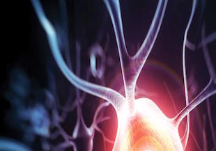
While the process of physiological nociception and pain perception is very complex, depending on the quality, intensity, and locality of the stimulus and the species, developmental age, and psychological state of the subjects (i.e., stress level, anticipation, emotional state, etc.), the general pathway for transmitting pain information to the brain is well documented. Nociceptors are pseudounipolar neurons with unencapsulated peripheral terminals the skin, muscles, joints, or viscera; cell bodies residing in the dorsal root ganglion (DRG); and central terminals in the dorsal horn of the spinal cord. There are generally two types of nociceptors – A‐fibers are fast‐conducting with myelinated axons and have small receptive fields for stimulus localization while C‐fibers are slower with unmyelinated axons that are bundled into fascicles wrapped by Schwann cells and have broad receptive fields. Nociceptors normally are electrically silent and have a high threshold compared to somatosensory neurons involved in, for example, vision or hearing. Once stimulated, nociceptors produce all or nothing action potentials releasing glutamate as their primary neurotransmitter and having excitatory effects on postsynaptic cells in the dorsal horn. In the dorsal horn, primary afferent neurons either synapse directly with projection neurons or, more commonly, first with a variety of excitatory and inhibitory interneurons for signal modification. Ascending projection neurons extend, mostly contralaterally, to supraspinal targets including the caudal ventrolateral medulla, the nucleus of the solitary tract, the lateral parabrachial area, the periaqueductal grey matter, and the thalamus. Descending pathways projecting from the nucleus raphe magnus and the locus coeruleus release serotonin and norepenephrin, respectively, via volume transmission in the DRG to further modify pain processing. All along the pain processing pathway, from the primary afferent nociceptors, to the dorsal horn of the spinal cord, to the supraspinal processing centers and including descending projections that further modify processing, there is a delicate balance of excitation and inhibition that is important for properly representing the pain stimulus. Miss‐communication at any of these locations can result in chronic pain.
Stay tuned as we continue to explore in the coming weeks Neuropathic pain, Cell Types involved in the Neuro-inflammation aspect or Neuropathic pain, Relevant preclinical models, and Potential inflammation-related drug targets for treatment of Neuropathic pain.
About MD Biosciences
MD Biosciences is a preclinical CRO offering services in neurology and pain. If you would like to speak with a scientist, please contact us now.