


4 min read
Histological Markers of Myelination in Demyelination Studies
By: MD Biosciences on Nov 3, 2026, 9:00:03 AM
Remyelination trials in multiple sclerosis are judged on imaging and functional endpoints because myelin cannot be examined directly in a patient. A preclinical study has that option, because tissue can be sectioned, stained and quantified, and the result can be related to the electrophysiology and behavior recorded in the same animals. Preclinical work is therefore the only stage of a remyelination program at which the intended myelin effect can be verified against the myelin itself.
The stains and markers used for this purpose address different questions, and a study designed to demonstrate remyelination usually requires several of them. MD Biosciences runs the panel described below across its central and peripheral demyelination and nerve repair models.
Myelin Basic Protein as the Direct Measure of Myelin
Myelin basic protein is a major structural component of the myelin sheath, and immunostaining for MBP is the most direct histological readout of myelin present in a section. In the cuprizone model, brains are sectioned at the corpus callosum and stained for MBP. The endpoints are MBP optical density within the corpus callosum and corpus callosum thickness. In the example data, cuprizone-fed mice show reduced corpus callosum staining at 6 weeks and more pronounced demyelination at 13 weeks, and corpus callosum width is reduced relative to naive controls at both terminations. Because cuprizone withdrawal allows spontaneous remyelination, the same MBP readout applied during the recovery phase quantifies the rate and extent of myelin restoration.
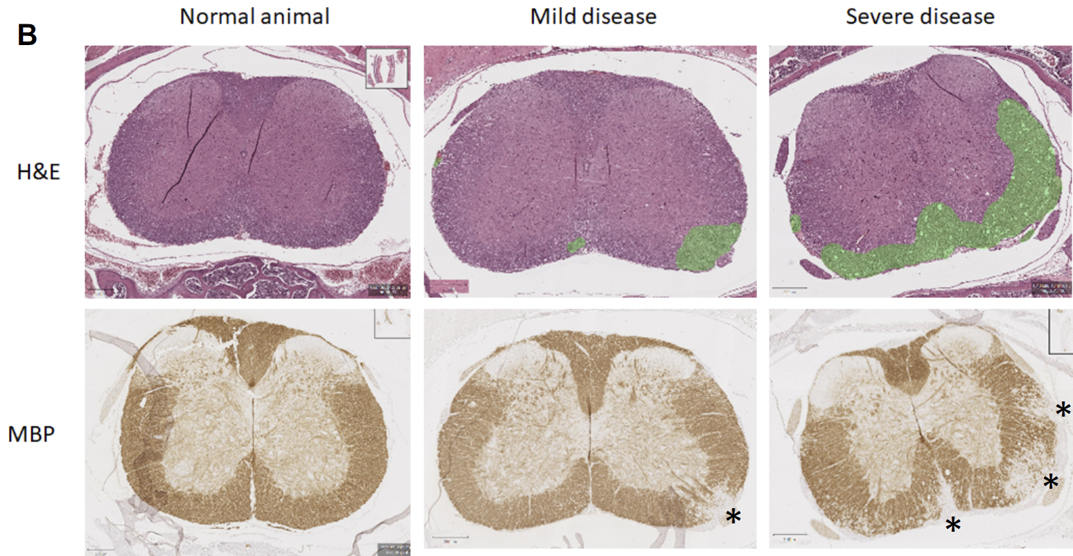
In MOG-induced EAE, MBP staining of longitudinal spinal cord sections distinguishes normal white matter, which stains dark brown against paler gray matter, from regions of white matter damage. The example sections show myelin debris within lesions and, at high magnification, myelin sheath fragments undergoing phagocytosis by macrophages in digestion chambers. Sections are taken at defined levels of the cord, with transverse sections at cervical, thoracic and sacral levels and longitudinal sections between them. Each level is scored so that lesion burden can be mapped along the length of the cord.
H&E Staining and the Cellular Context of the Lesion
Hematoxylin and eosin staining shows the cellular environment surrounding the myelin loss. In MOG-EAE spinal cord, H&E reveals multifocal vacuolation in the ventral white matter with myelin debris within some vacuoles, increased cellularity from gliosis and inflammatory infiltration, hypercellular foci, and meningeal inflammation. Evaluated alongside MBP staining of the same lesion, H&E indicates whether an area of myelin loss lies within active inflammation or represents residual damage after inflammation has resolved. This distinction defines the lesion environment in which a remyelinating candidate is intended to act.
Glial Markers of the Cellular Response
Demyelination and repair are cellular events, and immunohistochemistry for the cells involved adds information that myelin stains alone cannot provide. GFAP marks astrocyte activation and Iba-1 marks microglia, so together they characterize the glial response around a lesion. MD Biosciences applies both across its neurodegeneration and neuroinflammation models, in paraffin or frozen sections, and the same antibodies transfer directly to a demyelination study.
Axonal Markers of Axon Survival
Myelin loss and axonal loss are separate outcomes, and a repair study must distinguish between them. Neurofilament staining, using NF-M or NF200, marks the axon itself and identifies the large myelinated fibers, and beta-III tubulin serves as a pan-neuronal axon integrity marker. In a demyelinating lesion, preserved neurofilament staining within an area of MBP loss indicates demyelinated but intact axons, which are the substrate on which remyelination acts. Where axons have degenerated, remyelination is no longer the relevant endpoint.
Peripheral Nerve Markers: Schwann Cells and Myelin Thickness
Peripheral remyelination is assessed with a related but distinct panel. S100b marks Schwann cells and indicates recolonization of a repair site, since Schwann cells produce peripheral myelin and their return precedes remyelination. The G-ratio, the ratio of axon diameter to total fiber diameter, quantifies myelin thickness relative to the axon and is the standard measure of remyelination quality in peripheral nerve repair. Total axon count and density across a repair site quantify regeneration, motor endplate staining demonstrates reinnervation of muscle distal to the injury, and distal muscle fiber size indicates whether denervation atrophy has been reversed. MD Biosciences runs this panel in its rat and Göttingen minipig nerve repair models. The peripheral nerve injury material includes H&E, S100b and MBP staining of the injured nerve together with intraepidermal nerve fiber counts in the foot.
Correlation of Histology With Function
Histology is terminal, so the functional readouts collected during the study connect it to the living animal. In MOG-EAE, transcranial motor evoked potentials degrade as clinical score rises, and visual evoked potential latency is lengthened by day 14 after induction, so conduction slows as demyelination develops. In peripheral nerve models, compound muscle action potentials and sensory nerve action potentials measure conduction across the repair site over months, and behavioral testing quantifies functional performance. When the histology at termination is interpreted against those longitudinal measures, a study can demonstrate not only that myelin was preserved or restored but that conduction and function improved concordantly.
Quantification is computational where the endpoint requires it. MBP optical density and corpus callosum thickness in the cuprizone model are measured rather than graded. MD Biosciences applies AI-based image analysis to CD31 vessel quantification, Herovici collagen staining and intraepidermal nerve fiber density, yielding continuous rather than ordinal measures.
MD Biosciences Capabilities
The full panel runs in house, with H&E performed alongside a validated antibody panel of MBP, GFAP, Iba-1, NF-M, NF200, beta-III tubulin, S100b, CGRP, synaptophysin, ChAT, NeuN, TH and active caspase 3 in paraffin or frozen sections. Additional markers can be validated on request, and computational quantification is available for the endpoints that benefit from it. A remyelination study can therefore include the myelin stain, the cellular context, the axonal status and the functional correlate in the same animals. Study design conversations are welcome at neuro@mdbiosciences.com.