


4 min read
Cuprizone and EAE Models of Demyelination: Mechanism and Application
By: MD Biosciences on Oct 13, 2026, 9:00:05 AM
Multiple sclerosis therapy has been transformed by agents that control inflammation, and the unmet need has shifted toward repair. Remyelination has proven a difficult target in the clinic. A small randomized trial of clemastine, a first generation antihistamine with muscarinic antagonist activity that was repurposed as a remyelinating agent, was the first to document a remyelination signal for a putative repair agent. The phase 2 VISTA trial of PIPE-307, an M1 receptor antagonist, did not meet its primary endpoint in relapsing MS despite being safe and well tolerated. Recent reviews of the field devote as much attention to trial design as to mechanism, because a remyelinating effect must be separated from anti-inflammatory effects before it can be measured.
That separation begins preclinically. The two most widely used demyelination models, experimental autoimmune encephalomyelitis and the cuprizone model, produce myelin loss through different mechanisms, and each is suited to a different type of question. MD Biosciences runs both models.
EAE: Demyelination Through Autoimmune Inflammation
Experimental autoimmune encephalomyelitis is the non-clinical model of autoimmune inflammatory disease of the CNS and resembles many aspects of human multiple sclerosis. Immunization with a myelin antigen produces an encephalitogenic T cell response. Pertussis toxin is co-administered to open the blood brain barrier, inflammatory cells enter the CNS, and demyelination follows. Because the immune response is primary, the model is used for therapeutics that act on the immune system or on the neuroinflammatory cascade, and for neuroprotective candidates evaluated against an inflammatory background.
The variant selected determines the disease course. MOG-induced EAE in C57Bl/6 mice produces a chronic progressive disease, with clinical signs appearing around day 10 and peaking around day 16 over a 21 to 25 day study. PLP-induced EAE in SJL mice produces a remitting and relapsing course over 50 days, with a first peak at days 14 to 16 and a relapse at days 25 to 30. The relapse is driven by epitope spreading, as T cells react to secondary myelin peptides released during the first phase. MBP-induced EAE in Lewis rats produces a single self-limiting episode of hind paw paralysis between days 10 and 21, with mononuclear cell infiltration but without demyelination. This variant therefore models the inflammatory event in isolation and provides a 21 day design for assessing an acute anti-inflammatory effect.
Disease is followed on a 0 to 5 clinical score alongside body weight and rotarod performance, with FTY720, dexamethasone and Copaxone as reference compounds. Electrophysiology adds a quantitative in vivo measure of conduction. In the example data, transcranial motor evoked potentials recorded from the tibialis anterior are desynchronized in mice at clinical score 1 to 2, with multiple peaks, delayed latency and extended duration. At score 3 to 4 the potentials are markedly reduced in amplitude. Spinal cord histology by H&E and MBP staining demonstrates the inflammatory lesion and the myelin loss within it. Cytokine profiling by multiplex analysis and pharmacokinetic sampling complete the study package.
Cuprizone: Demyelination Without a Primary Immune Response
The cuprizone model produces demyelination without an immune trigger. Cuprizone, a copper chelator administered in the diet, induces oligodendrocyte death in a reproducible pattern, with the corpus callosum the most affected region. No primary autoimmune response is involved, and after withdrawal of the diet the corpus callosum remyelinates spontaneously. The model therefore isolates the oligodendrocyte and the myelin sheath as the objects of study, which suits neuroprotective and remyelinating therapies whose mechanism is independent of the immune system.
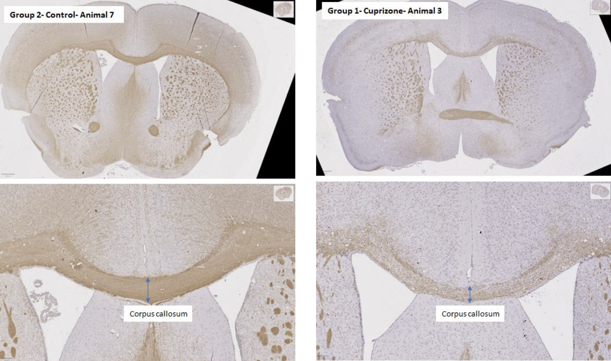
The MD Biosciences protocol uses male C57BL/6 mice of 8 to 10 weeks, with rapamycin co-administered with the cuprizone diet so that demyelination develops consistently across animals. Studies run 7 to 13 weeks and body weight is tracked weekly. At termination the brain is sectioned through the corpus callosum and stained for MBP, with MBP optical density and corpus callosum thickness as the endpoints. In the example data the corpus callosum is narrower in cuprizone animals than in naive controls at both terminations. The loss of MBP staining is more pronounced at the 13 week timepoint than at the earlier one.
Applications of Each Model
The two models are complementary. EAE is used to assess whether a candidate alters the course of an immune-mediated demyelinating disease, measured by clinical score, conduction, histology and cytokines. Because its demyelination is a consequence of inflammation, a candidate that reduces demyelination in EAE may act by suppressing the immune attack rather than by protecting oligodendrocytes or promoting repair.
The cuprizone model is used to assess whether a candidate protects oligodendrocytes or accelerates remyelination in the absence of an immune driver. A candidate that preserves MBP density or corpus callosum thickness in cuprizone animals acts on myelin biology directly. A candidate that accelerates recovery after cuprizone withdrawal demonstrates a remyelination effect that EAE cannot isolate.
A remyelination program typically requires both models. The cuprizone model establishes that the mechanism acts on oligodendrocytes and myelin, and EAE establishes whether that effect persists in the inflammatory environment characteristic of the patient population. The PLP relapsing model addresses a further question, whether a repair effect is maintained across cycles of inflammation and remission.
MD Biosciences Capabilities
Both models run in house, with MOG, PLP and MBP variants of EAE and the rapamycin-supported cuprizone protocol. Across them, MD Biosciences applies clinical scoring, rotarod, transcranial motor and sensory evoked potentials, visual evoked potentials, H&E and MBP histology of spinal cord and brain, immunohistochemistry for cellular infiltration, and multiplex cytokine analysis. A program can advance a candidate from the cuprizone model into an EAE variant using the same histological readouts, so that the remyelination result and the disease modification result are directly comparable. Study design conversations are welcome at neuro@mdbiosciences.com.
References:
The Road to Remyelination in Multiple Sclerosis: Breakthroughs, Challenges, and Considerations for Future Trial Design. Drugs, 2025.
Cuprizone and Experimental Autoimmune Encephalomyelitis models. MD Biosciences datasheets.