

2 min read

Neurons communicate with each other as well as other muscles and organs through electrical events called action potentials and neurochemical transmitters. A synapse is the junction between two neurons where an action potential will cause one neuron to release a neurochemical transmitter that will either excite or inhibit the adjacent neuron from firing its own action potential.
These action potentials are the main means of communication between neurons. In an intact brain, it is the balance of these inhibitory and excitatory inputs to a neuron that determines if an action potential will occur.
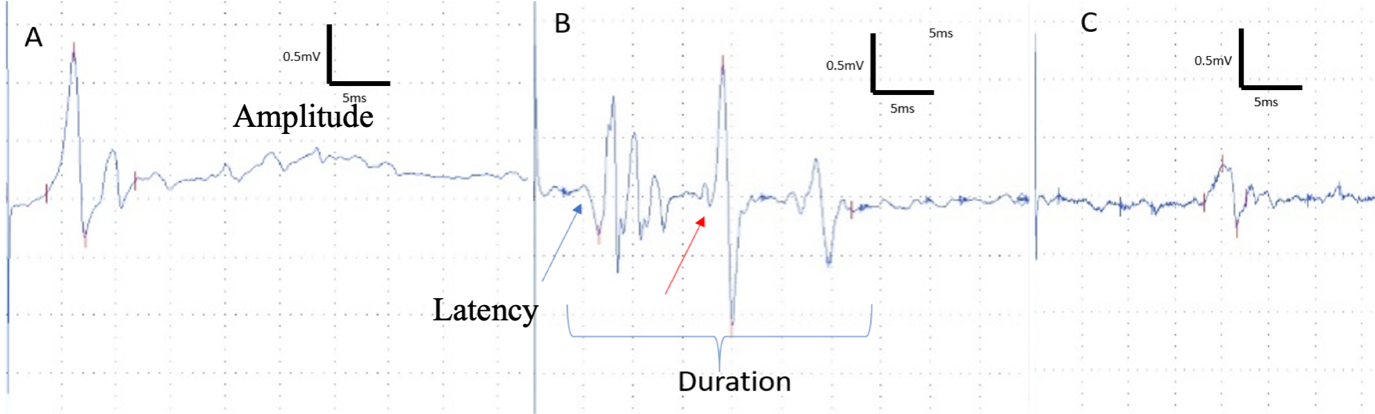
Using electrophysiology, we are able to study these neuronal signals in pain and CNS disorders. Evoked potentials (EP) measure the electrophysiological responses of the nervous system to external stimuli and provide information on both peripheral and central nervous system pathways. Evoked potential responses consist of a sequence of peaks and waves that are characterized by latency, amplitude, and intervals between peaks.
Measuring evoked potentials in animal models provides an objective physiological marker of clinically relevant abnormalities in the sensory or motor systems. Sensory-evoked potentials (SEPs) are used to increase knowledge about nociception and pain. SEPs evoked by high intensity stimuli are believed to reflect the processing of noxious stimuli, correlating well with subjective pain ratings in the clinical situation, the instance of pain in animals, and the alterations observed by therapeutic treatments in both the clinical and preclinical situation. SEPs are therefore considered to be a potential method for quantifying acute pain as well as differentiating the sensory and affective component of acute pain.

Motor evoked potentials (MEP) are neuroelectrical signals that are produced by the spinal cord or peripheral muscles and provide an objective assessment of motor function integrity. Stimulation of the motor cortex produces a response in a targeted muscle and the latency of the response indicates the time for the impulse to reach that targeted muscle. It has been shown to have good correlation to neurological deficits in both human and animal.
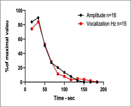
Measuring both SEPs and MEPs, we are able to measure nerve regeneration, degeneration and protection in response to various treatments. For example, in EAE studies for MS, we are able to measure pain in animals that have limited mobility by correlating vocalization to SEP amplitude.
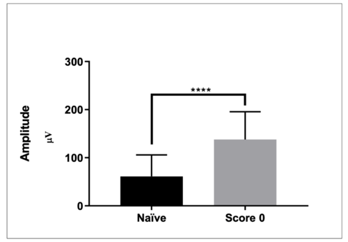
SEP readings also show changes prior to motor dysfunction. This suggests that onset of disease may begin prior to what is seen in clinical scores, providing insight into when therapeutic treatment should start for greater efficacy.
The field of pain and CNS research has advanced tremendously over the past decade, however is still challenged by low drug approval rates compared to other therapeutic areas. In vivo electrophysiology is a clinically relevant readout that allows us to study the function and connectivity of neurons, reveal abnormalities, and assess whether drug candidates can restore normal neural function.
Interested in how your preclinical study would benefit from electrophysiology monitoring?

Related Posts
Importance of Diversity in Preclinical Stroke Models
The Need for Diversity in Animal Models
The clinical population is diverse, including both sexes,...
Increase diversity to improve clinical translation and strengthen pain management outcomes.
The goal of preclinical drug development is to identify potential therapeutic compounds and gain an...
Behavioral Research Assessments & Applications: Quantify Cognitive, Emotional, Sensory & Motor Functions
Neurological diseases often result in a combination of motor and cognitive deficits. Thus,...