

4 min read
Biomarker Strategy in Large Animal Preclinical Studies: What Actually Translates
By: MD Biosciences on May 19, 2026 9:00:03 AM
Biomarkers have become the connective tissue of modern drug development. They anchor mechanism of action claims, support dose selection, enable patient stratification, and in many therapeutic areas now serve as the primary efficacy readouts in early clinical trials. For preclinical programs, that shift has raised the stakes on biomarker strategy in a specific way. It is no longer enough to measure something. The measurement has to translate.
In small animals, biomarker selection is often driven by pragmatism. What can we measure in this species, at this sample volume, with this analytical pipeline? In large animal studies, the calculus changes. Blood volumes allow repeat sampling, tissue access permits serial biopsies, and the physiology supports panels of biomarkers that map more directly onto what clinicians measure in patients. Designing those panels well is where a meaningful fraction of translational value gets created, or lost.
The Three Biomarker Categories That Matter Most
Across therapeutic areas, preclinical biomarker panels in large animals tend to fall into three categories, and the best programs integrate all three rather than treating them as separate deliverables.
Circulating biomarkers are the most portable. Blood based measurements of neurofilament light chain (NFL) for neurodegeneration, KIM 1 and NGAL for kidney injury, troponin and CPK for cardiac injury, and a range of inflammatory cytokines for immune activation all have established clinical utility. Because the analytical methods and the underlying biology travel well between species, circulating biomarkers are often the primary translational tool in a large animal package. The key question is not whether to measure them, but which specific analytes correspond to the biology the program is trying to perturb.
Tissue biomarkers are where large animal studies begin to differentiate themselves. In a well designed porcine pain study, skin biopsies from the same animal that provided behavioral and electrophysiology data can be interrogated for intraepidermal nerve fiber density using anti PGP9.5, for neuropeptide expression (CGRP, substance P), for sodium channel localization (Nav1.7), and for endothelin receptor distribution (ETA, ETB). This is the endpoint layer where the case for porcine models becomes most compelling, because the histological findings in pig skin mirror those in human neuropathic pain biopsies (Rice et al., 2019, Neurobiology of Pain), including the CGRP and Nav1.7 increases and the ETB decrease characteristic of human disease.
Functional biomarkers sit at the interface of physiology and behavior. Electrophysiology readouts, including sensory nerve conduction velocity, compound muscle action potentials, and sensory nerve action potentials, have long been standard in clinical neurology. MD Biosciences pioneered the first cMAP and SNAP recordings in swine, extending a clinical measurement technique into a preclinical species with comparable nerve architecture. These functional biomarkers are not substitutes for circulating or tissue biomarkers. They are orthogonal measurements that, when combined with the other two categories, triangulate the biology in ways that no single readout can.
What Neurodegeneration Programs Should Be Measuring
Neurodegenerative disease programs have been ahead of other therapeutic areas in biomarker sophistication, and the panel that has emerged is informative for large animal design. Neurofilament light chain captures axonal damage and has clinical utility across multiple sclerosis, Alzheimer's disease, Parkinson's disease, ALS, and traumatic brain injury. GFAP tracks astrocyte activation and neuroinflammation. Alpha synuclein is a Parkinson's specific readout where assay maturity is advancing. Tau, in its various phosphorylation and cleavage states, supports Alzheimer's and tauopathy programs. BDNF serves as a neurotrophic readout relevant to multiple CNS indications.
In an MD Biosciences multiple sclerosis model, these circulating biomarkers can be integrated with tcMEP and tcSEP electrophysiology, clinical scoring, and cytokine multiplex panels including IL 1 beta, TNF alpha, IFN gamma, IL 10, and others. The novel P25 sensory evoked potential wave, identified and characterized by MD Biosciences researchers in MOG induced EAE (Shulman et al., Journal of Pain), illustrates what becomes possible when functional biomarkers are pursued specifically rather than generically. The P25 wave increases in early mild EAE as a signature of hypersensitivity, correlates with vocalization, responds to morphine, and disappears in severe late stage disease. That kind of quantitative in vivo biomarker, tied to a defined disease stage and a defined therapeutic response, is what sponsors building MS programs increasingly want.
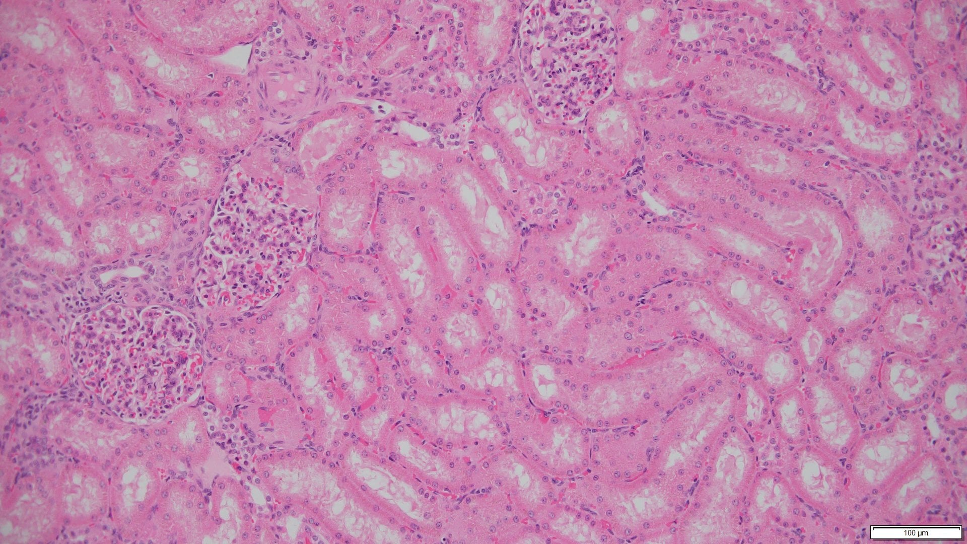
Kidney, Cardiac, and Wound Healing Panels
Outside the CNS, biomarker strategy in large animals follows similar logic. Kidney studies integrate blood creatinine, BUN, KIM 1, and NGAL with urinary protein and albumin, alongside histopathology that maps onto human clinical pathology. Cardiac studies pair CPK and troponin with echocardiography and histology. Wound healing studies combine cytokine multiplex (TNF alpha, IL 8, MCP 1, VEGF A) with quantitative histology for granulation tissue thickness, collagen phenotype via Herovici staining, and blood vessel density via CD31.
The common thread is that the biomarkers selected are the same ones clinicians use, or the same analytes measured using methods that translate cleanly to clinical assays. That is the structural difference between a preclinical biomarker panel designed for translation and one designed for publication.
AI Histology as an Enabling Layer
Much of what makes a large animal biomarker panel useful depends on the quality of the histology. Manual quantification has historically been the rate limiting step, both in throughput and in reproducibility. MD Biosciences uses an AI platform to quantify CD31 for angiogenesis, Herovici staining for collagen typing, and PGP9.5 for intraepidermal nerve fiber density. The ChemoMorphometric Analysis (CMA) platform extends this to proprietary multi antibody skin biopsy workflows that produce integrated quantitative outputs.
The operational benefit is throughput. The scientific benefit is reproducibility. Across multiple studies, across multiple technicians, a quantitative histology platform produces outputs that can be compared, pooled, and interrogated in ways that manual scoring cannot support at scale.
How to Build a Biomarker Strategy That Actually Serves the Program
The strategic error that appears most often in preclinical planning is treating biomarkers as a downstream deliverable rather than a design input. The most effective programs decide early which clinical biomarkers will define the Phase 1 or Phase 2 trial, and then build backward into the preclinical package to ensure those biomarkers are measured in a relevant species using methods that will support cross study comparison.
For programs running large animal studies, the questions worth asking at the planning stage are specific. Which clinical biomarkers will matter in the patient population? Which of those biomarkers translate across species, and which require species adaptation? Which tissue biomarkers can be assessed in the same animal providing circulating and functional data? And what analytical methods will produce data that the clinical program can actually use when the time comes?
A biomarker strategy built on those questions, rather than on what is easy to measure, is the one that creates the strongest translational bridge.
— — —
MD Biosciences provides integrated biomarker development across pain, neurodegeneration, wound healing, kidney, and cardiac programs, with circulating, tissue, and functional readouts quantified through AI histology and validated assay platforms. For questions about biomarker strategy, contact neuro@mdbiosciences.com.
Related Posts
The Diabetic Wound Healing Model That Keeps Earning Its Keep
Chronic wounds in diabetic patients are one of the costliest quietly growing burdens in medicine....
CIPN Keeps Failing in the Clinic. The Preclinical Package May Be Why.
Chemotherapy induced peripheral neuropathy remains one of the most underserved symptoms in oncology...
Stroke Models Need Diversity: What Age, Sex, and Comorbidity Change About the Data
Stroke remains one of the highest attrition areas in drug development. Despite decades of...