

10 min read
Key features of the 6-OHDA model, their benefits and drawbacks
By: MD Biosciences on Jun 2, 2013 10:01:00 PM
The 6-OHDA model has contributed significantly to our understanding of Parkinson’s Disease and the development of treatments. Like every animal model, there are limitations to recapitulating human disease. This review will discuss some of the benefits and drawbacks of the 6-OHDA model of PD.
6-OHDA is injected directly into the brain
Because 6-OHDA does not cross the blood-brain barrier, it cannot be dosed systemically and must be injected stereotaxically directly into the brain. This requires surgical instrumentation, facilities and training in their use. Even so, the procedure is of relatively low complexity and cost, and the 6-OHDA-induced lesion is highly reproducible. Injections are typically made into one of three general locations: the substantia nigra pars compacta (SNpc), medial forebrain bundle (mfb) or the striatum. Sometimes, specific sub-regions within these areas are targeted, such as only the A9 fibers in the mfb [21], or specific areas of the striatum (e.g. dorsolateral, ventrocentral, etc.) [14]. All injection locations eventually result in a loss of dopaminergic input to the striatum.
6-OHDA is relatively specific for catecholaminergic neurons.
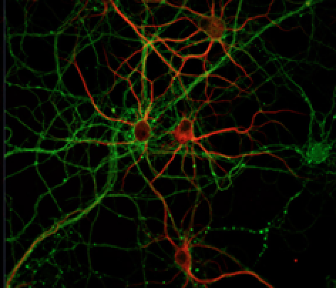
6-OHDA has high affinity for the catecholamine transporters due to its chemical similarity with the catecholamines. Following injection, it is taken up into the cytosol of dopaminergic neurons by the dopamine transporter. The model produces degeneration and death of a significant percent of dopaminergic neurons on the SNpc and fibers in the nigrostriatal tract, and reduces the amount of dopamine in the SNpc and the striatum, as assessed by immunostaining for tyrosine hydroxylase, a key enzyme in the dopamine synthetic pathway.
The similarity of 6-OHDA to the catecholamines, however, also means that 6-OHDA has a high affinity for the transporters for other catecholamines, such as noradrenaline and serotonin, and that it is toxic to these neurons as well. To prevent these other catecholaminergic neurons from being damaged when using this model, and to improve specificity of 6-OHDA for dopaminergic neurons, a selective noradrenaline reuptake inhibitor, such as desipramine, is often administered systemically 30-60 minutes before 6-OHDA. (But since PD patients are known to also lose noradrenergic neurons, in the locus coeruleus, some argue that adding desipramine reduces the model’s translational value.) Pargyline, a monoamine oxidase inhibitor which prevents the metabolism of 6-OHDA, may also be given prolong the 6-OHDA effect or minimize the dose required.
6-OHDA is usually injected unilaterally, resulting in a hemi-parkinsonism model.
A unilateral model is almost always used because bilateral injections result in adispsia, aphagia, seizures and inability of rats to care for themselves. In the unilateral model mortality is typically not an issue. Some of the tests of non-motor function, however, have used bilateral injections, as noted later in this article.
A key benefit of the unilateral model is that each animal can serve as its own control (there is a lesioned and a non-lesioned hemisphere), and the extent of degeneration can be measured post-mortem by comparing lesioned and intact hemispheres. The downside of the unilateral model, and the unilateral symptoms it produces, is that it does not mimic human PD, which is bilateral.
The model produces rapid results, and stable histology and behaviors.
6-OHDA injections, especially into the SNpc and mfb, produce a rapid degeneration, beginning in 12-24 hours, and maximal around 1 week, though details vary somewhat between reports. The time course of degeneration following striatal injections is slower, but still rapid compared to the multi-year degeneration in human PD. Both models yield a static disease state when lesions are large, although partial recovery at 2-3 months post-injection can be observed in the partial-lesion models, and this can complicate results of behavioral testing. Spontaneous motor behaviors e.g. forelimb akinesia, impairment in initiating stepping movements, preference for using the non-impaired forelimb, are observed, and many are reversed by L-dopa. Impairment in non-motor symptoms also has been observed, although with less consistent results (reviewed in 3, 9, 16, 17].
The main characteristics of PD that are not recapitulated by the 6-OHDA model are Lewy bodies (the main histopathological hallmark of the disorders), resting tremor, and a slow, progressive disease with neuronal degeneration on-going for years. Findings are inconsistent as to whether the model captures the clinical PD features of pathology in other brain areas e.g. the locus coeruleus and raphe nuclei, or involvement of other transmitter systems, e.g. the serotonergic system.
Complete or partial lesions of the SNpc can be produced.
The 6-OHDA model can be tuned to mimic different stages of the PD disease process. Partial lesions are thought to better resemble earlier stages of PD, i.e. before the appearance of, or at the onset of motor symptoms. In human PD, it is believed that motor symptoms do not appear until approximately 80% of the dopaminergic neurons in the SNpc have died, and non-motor symptoms are thought to become apparent with cell loss around 50%. Earlier disease states are thought to be modeled by injections in the striatum (or by low dose infusions into the SNpc or mfb), and are appropriate for studying neurodegenerative processes or neuroprotective strategies. End-stage disease states are produced by lesions of the SNpc or mfb, and are useful for assessing efficacy of dopamine replacement therapies or cell transplants, or effects of long-term use of L-dopa. The behavioral changes following striatal injection, or low dose injections in the SNpc or mfb, appear to be more variable than after full lesions [10].
Three sites of dosing and the outcomes that can be expected.
The three most common sites for intracerebral 6-OHDA infusion are the medial forebrain bundle (mfb), the substantia nigra pars compacta (SNpc), and the striatum. The choice of injection site can be influenced by a number of factors:
- The question being pursued - is the investigator interested in downstream consequences of nigral cell death, cell replacement therapies, dopaminereplacement therapies, neuroprotection?
- Practical considerations or experimental needs, e.g. to not cause mechanical damage to the injection site, or to prepare for later experimental phases
- The extent of damage the investigator is trying to induce - SNpc and mfb injections typically produce more damage than striatal injection.
- Speed of result - SNpc and mfb injections result in more rapid neurodegeneration than striatal injection
- Profile of cell death - nigral neurons are affected first with SNpc dosing; striatal terminals degenerate first with striatal injection
Striatum
The striatum (the caudate-putamen complex) is the subcortical structure into which the terminals of the SNpc neurons project. The method of injecting 6-OHDA into the striatum, either as a single injection or as multiple injections into multiple striatal locations, is often the choice for investigating mechanisms of PD pathogenesis and cell death, or testing neuroprotective strategies. Striatal injections, and the retrograde degeneration they produce, are also sometimes chosen because the pattern of degeneration in this model - striatal terminals degenerating before TH-positive neurons in the SNpc is believed to mirror better the clinical disease. However, even with this more slowly evolving 6-OHDA approach, cell death and dopamine depletion still occur quite rapidly compared to human PD, and there is considerable concern that even this slower version of the 6-OHDA model may not be able to accurately model the extremely slow and progressive chain of events that eventually leads to human PD.
Outcome: Injection of 6-OHDA into the striatum causes a slowly developing, partial lesion of the nigrostriatal pathway, with retrograde degeneration of dopaminergic neurons in the SNpc, and loss of dopamine activity in the striatum. The striatal model is usually considered to be a “partial lesion” model. Partial lesion models often aim to produce 60-70% degeneration of the nigrostriatal tract because this is the level at which motor symptoms of PD appear. With most partial lesion approaches the extent of cell loss in the SNpc achieved varies among laboratories. For example, injection of 20-28 ug 6-OHDA has resulted in 20-85% loss of SNpc cells, and a 60-90% reduction in striatal dopamine [reviewed in 10].
Time course: A single striatal infusion of 6-OHDA (total dose of ~20-30 ug) reduces TH staining by about 33% within 24 hours. The lesion size increases to cover a maximum of about 66% of the injected striatum over the following 2-3 weeks [1, 2]. In the SNpc, cell loss is minimal at 1 week, reaching a maximum at around 2-3 weeks post injection. The loss of TH immunoreactivity continues to a maximum of 50-60% of the nucleus within 3-4 weeks [1,2] As noted above, the specific details vary among laboratories and depend on the exact details of the experiment, including the specific location of the striatal injection [14].
Histopathology: Dying nigral neurons exhibit a heterogeneous morphology, including apoptotic-like features. The pattern of cell loss mirrors that observed in PD, with about 20% greater loss in the SNpc as compared to the ventral tegmental area. An inflammatory response also has been reported with striatal infusions: activated microglia can be detected in both the SNpc and the striatum at about 1 week post-injection [6, 10].
Medial forebrain bundle injection (mfb)
The mfb contains the axons of the dopaminergic nigral neurons which project to the striatum. Injection of 6-OHDA unilaterally into the medial forebrain bundle (mfb) is the model that has contributed most to preclinical research on PD. Infusion into the mfb is suitable for studying the consequences of dopaminergic cell death and for testing strategies to treat motor symptoms and treatment-induced dyskinesias [reviewed in 4]. This site of dosing might be selected when it is desired to prevent direct damage to the SNpc or minimize undesired effects on neurons in the ventral tegmental area, located just ventral to the SNpc, and to which toxin injected into the SNpc can spread.
Outcome: Injection of 6-OHDA (~16 ug) into the mfb unilaterally can cause a total or near total destruction of dopaminergic neurons in the SNpc (the A9 cells) and the ventral tegmental area, VTA (A10 cells). This results in a significant (>80%) depletion of dopamine in the ipsilateral striatum, and in denervation supersensitivity of the postsynaptic dopamine receptors on the injected side of the brain [9, 21].
Time course: There is some indication that neuronal degeneration and dopamine loss in the mfb model may proceed differently than in the direct SNpc model. Three weeks after lesioning (the earliest time point examined), denervation of the striatum was nearly complete, with a 99% decrease in dopamine content over the intact hemisphere. However, in the SN at the same time, the number of tyrosine hydroxylase immunoreactive (TH-IR) cells had only dropped to 88%. TH-IR continued to decline, to a 99% loss when examined 2 weeks later [24].
Histopathology: By some accounts, degenerating neurons in the SNpc take on a non-apoptotic morphology [13], whereas others report increased TUNEL staining, indicative of apoptosis, in the ventromedial region of the SNpc at 6, 48 and 96 hours [30].
Dosing: Some groups have been able to generate graded lesions, in which cell loss correlated with dose of toxin injected. Doses of 4µg, 8µg and 16µg of 6-OHDA resulted in cell loss (as measured by TH-IR) of 20%, 80% and 95%, respectively [27]. And injections of 6µg into the mfb resulted in 65% cell loss in the SNpc [28].
Substantia nigra pars compacta injection (SNpc)
The substantia nigra is the midbrain nucleus where the cell bodies of dopamine producing neurons are located. Injecting 6-OHDA directly into the SNpc is suitable for studying the consequences of dopaminergic cell death, and for testing strategies to treat motor symptoms associated with the disease, and dyskinesias produced by dopamine replacement therapy.
Outcome: Injection of high amounts of 6-OHDA into the SNpc can produce a rapid and virtually complete (80-100%) loss of nigral dopaminergic cell bodies. Toxic effects have been shown to spread to neurons in the ventral tegmental area (VTA), causing 40% loss of cells in that area [8]. Cell loss in the SNpc is followed by degeneration of the vast majority of dopaminergic terminals in the striatum (the area to which the SNpc dopaminergic neurons project) and depletion of striatal dopamine, as measured by tyrosine hydroxylase immunoreactivity. Time course: Following SNpc infusion, cell death in the SNpc begins as early as 12 hours [13] and typically within 24 hours; in the striatum, dopamine content becomes reduced below that of the uninjected side by about 3 days and continues to fall up to 10 days later, and remains stable for at least 4 weeks post lesion.
Histopathology: Degenerating neurons in the SNpc take on a non-apoptotic morphology [13]. The pattern of dopaminergic cell loss mirrors that seen in PD, where loss of cells in the SNpc (the “A9” cells) is more extensive than in the Ventral Tegmental Area (the “A10” cells).
Dosing: Some groups have been able to inject 6-OHDA in a dose-dependent manner to generate a partial lesion which may better resemble earlier stages of PD, e.g. when motor symptoms are not apparent. For example, an injection of 10 ug produced almost complete depletion of dopamine in the SN, and 6 ug decreased dopamine by 50% [7].
References:
- Blandini, F., Levandis, G., Bazzini, E., Nappi, G. & Armentero, M.-T. Time-course of nigrostriatal damage, basal ganglia metabolic changes and behavioural alterations following intrastriatal injection of 6-hydroxydopamine in the rat: new clues from an old model. The European journal of neuroscience 25, 397–405 (2006)
- Blandini, F., Armentero, M.-T. & Martignoni, E. The 6-hydroxydopamine model: news from the past. Parkinsonism & related disorders 14 Suppl 2, S124–9 (2007).
- Branchi, I. et al. Nonmotor symptoms in Parkinson’s disease: investigating early-phase onset of behavioral dysfunction in the 6-hydroxydopamine-lesioned rat model.Journal of neuroscience research 86, 2050–61 (2008).
- Bové, J. & Perier, C. Neurotoxin-based models of Parkinson’s disease. Neuroscience 211, 51–76 (2012).
- Chang, J., Wachtel, S., Young, D. & Kang, U. Biochemical and anatomical characterization of forepaw adjusting steps in rat models of Parkinson’s disease: studies on medial forebrain bundle and striatal lesions. (1999). at
- Cicchetti, F., Brownell, A.L., Williams, K., Chen, Y.I., Livni, E., Isacson, O. Neuroinflammation of the nigrostriatal pathway during progressive 6-OHDA dopamine degeneration in rats monitored by immunohistochemistry and PET imaging. European Journal of Neuroscience 15, 991-998 (2002).
- Costa G; Abin-Carriquiry JA; Dajas F Nicotine prevents striatal dopamine loss produced by 6-hydroxydopamine lesión in the substantia nigra. Brain research. 888(2). 336-342. (2001).
- Deumens, R., Blokland, A. & Prickaerts, J. Modeling Parkinson’s disease in rats: an evaluation of 6-OHDA lesions of the nigrostriatal pathway. Experimental neurology175, 303–17 (2002).
- Dunnett, S. & Lelos, M. Behavioral analysis of motor and non-motor symptoms in rodent models of Parkinson’s disease. Progress in brain research 184, 35–51 (2009).
- Duty, S. & Jenner, P. Animal models of Parkinson’s disease: a source of novel treatments and clues to the cause of the disease. British journal of pharmacology 164, 1357–91 (2011).
- Fleming, S., Schallert, T. & Ciucci, M. Cranial and related sensorimotor impairments in rodent models of Parkinson’s disease. Behavioural brain research 231, 317–22 (2012).
- Han, F. & Wang, H. Effects of desensitized nicotinic receptors on rotational behavior in a 6-hydroxydopamine model of Parkinson’s disease. Neuroscience letters 415, 200–4 (2007).
- Jeon, B., Jackson-Lewis, V. & Burke, R. 6-Hydroxydopamine lesion of the rat substantia nigra: time course and morphology of cell death. Neurodegeneration : a journal for neurodegenerative disorders, neuroprotection, and neuroregeneration 4, 131–7 (1995).
- Kirik, D., Rosenblad, C. & Björklund, A. Characterization of behavioral and neurodegenerative changes following partial lesions of the nigrostriatal dopamine system induced by intrastriatal 6-hydroxydopamine in the rat. Experimental neurology 152, 259–77 (1998).
- Lee, C., Sauer, H. & Bjorklund, A. Dopaminergic neuronal degeneration and motor impairments following axon terminal lesion by instrastriatal 6-hydroxydopamine in the rat. Neuroscience 72, 641–53 (1996).
- Lindgren, H. & Dunnett, S. Cognitive dysfunction and depression in Parkinson’s disease: what can be
Related Posts
Weekly Science Blog: Possible Therapies for Spinal Cord Injuries, Reversal for Inflammatory Diseases and More!
It's Tuesday, which means MD Biosciences is providing coverage of the latest fascinating and...
Preclinical Post-operative Pain Model Resembles Human Physiology
The management ofpost-operative painis a challenge for both physicians and patients. In addition...
A minipig Model of Incisional pain
The use of animal models for the research of post-operative pain (POP) has been well described[1]....