

11 min read
 Diseases of the central nervous system are extremely debilitating, increasingly common, and affect millions of people worldwide. Neurodegenerative diseases often result in a combination of cognitive and motor deficits that affect an individual’s ability to perform daily tasks. Affected patients become dependent on medical services and family support as their disease progresses. Improvements in health have lengthened lifespan; however, this has resulted in a higher incidence of neurodegenerative conditions that affect the aging population. Genetics often has a major role in the development of neurodegenerative diseases and disease severity sometimes increases with each generation. However, environmental factors may be equally important in contributing to the severity, progression, and outcome of patients with other types of neurodegenerative disorders. Exposure to environmental toxins, metals, industrial chemicals, and certain dietary or lifestyle factors, may result in damage to the nervous system. Acute brain injuries, such as head trauma or oxygen deprivation, can also result in cognitive impairments.
Although there are a variety of pathways leading to neurodegenerative disease, the end result is the same: loss of function in the central nervous system and resulting impairments. Many neurodegenerative disorders are progressive in nature, resulting in a decline in function that may occur over several years or several decades. While there are some treatments available for neurodegenerative disorders, there are no cures. Therefore, there is great interest in researching neurodegenerative diseases and a need to halt or slow the process of cognitive decline. Understanding the basic biological mechanism of disease and cognitive impairment is pertinent for the development of therapeutics.
The outcome in oxygen stress and neurodegenerative diseases
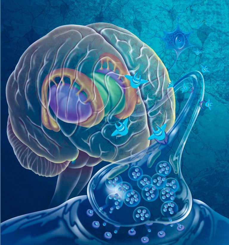
Homeostasis is critical for cell viability and cell maintenance. In cases of oxygen deprivation, autoimmune disease, or expression of disease factors, the cell is no longer able to maintain homeostasis and perform its necessary functions. Affected cells may die by necrosis or by apoptosis. When dead cells accumulate, reactive glial cells migrate to the site of injury and begin clearing the debris, but they also form scars that hinder axon regeneration and remylination [1]. Immune activation of glia may cause bystander effect and result in damage to the surrounding tissues. While it was once thought that cells of the central nervous system were unable to regenerate, we now know that restrictive microenvironmental factors are mostly responsible for preventing repair [2]. Strategies such as stem cell therapy, nanoparticle drug delivery, and gene therapy are being studied to determine if modulating the cellular microenvironment can lead to repair in the central nervous system [3-5].
Patients with cell death as a result of prolonged oxygen deprivation or disease have functional deficits. Although the young brain does display a high degree of plasticity, the adult brain is not easily able to regain lost function. After a stroke, the adult brain must undergo extensive changes in the motor cortical network in order to overcome resulting motor deficits. In patients with autoimmune disorders or neurodegeneration, the disease is typically progressive and patients are less likely to show any improvement in cognitive or motor function. Both cognitive function and motor function are typically affected by these progressive and acute brain injuries. Distinguishing between loss of motor function and loss of cognitive function is important for accurately studying these diseases and injuries in animal models.
Distinguishing between motor function and cognition
Differentiating between motor function and cognitive deficits can sometimes be a difficult task. For example, slow and slurred speech may lead a listener to suspect a patient has problems with language processing. However, difficulty with speech could be caused by damage to the hypoglossal nerve that controls movement of the tongue. Different areas of the central nervous system are responsible for voluntary (cognitive) and involuntary (reflexive) muscle movements. Deficits in purely motor function are often seen as problems in coordination, balance, and strength. A majority of involuntary motor function is controlled by the spinal cord. Even processes like walking are regulated by a feedback loop within the spinal cord and individuals do not consciously consider how to lift and place each leg. The cerebellum coordinates movement fluidity through feedback prediction. Patients with degeneration of the cells of the cerebellum have difficulty reaching for an intended target. Other motor deficits may lead patients to experience tremors, which are common in Parkinson’s disease. While lost motor functions are extremely problematic in performing daily tasks, some functions may be regained under certain conditions. In one study, 76% of patients regained motor functions six months after suffering a stroke [6]. Motor function was also regained in monkey models of brain lesions [7].
Cognitive regulation of motor function (motor planning) is controlled by the cerebral cortex. When an individual makes a decision to move, the cerebral cortex sends signals to the spinal cord where information is streamlined and relayed to muscles. Defects in cognitive planning may hijack successful completion of the motor task at hand. One of the paradoxes of Parkinson’s disease is that patients lose the cognitive decision to move, however the motor function needed to perform the action is maintained. Cognitive impairments do not appear to resolve as easily as motor impairments. The probability of long-term cognitive improvement after a stroke was only 54% for patients with damage to the left hemisphere of the brain [8]. When oxygen deprivation or neurodegenerative processes occur, patients often experience varying de- grees of loss of motor function combined with cognitive deficits. A stroke patient usually has muscle weakness on the contralateral side of the body and cognitive impairments related to the area of the brain that was damaged.
Because disease patients often experience varying degrees of motor and cognitive impairments, it is difficult to study neurodegenerative disease and cognition in humans. Similar biological processes control loss of cognitive function in humans and animals; therefore mouse models recapitulate the cell biology resulting in cognitive decline.
Mouse models provide many advantages to the study of neurodegenerative disorders. Models such as EAE for neuroimmunological disorders, the 4VO and MCAo for oxygen deprivation and 6-OHDA for neurodegenerative disease can be used to study cognitive impairments related to human disease.
 |
References:
-
Fawcett J.W., & Asher, R.A. The glial scar and central nervous system repair. Brain Research Bulletin. 49, 377-391 (1999)
-
Horner, P.J., Gage, F.H. Regenerating the damaged central nervous system. Nature 407, 963-970 (2000)
-
Berry, M., Barrett, L., Seymour, L., Baird, A., Logan, A. Gene therapy for central nervous system repair. Current Opinion in Molecular Therapies 3, 338-349 (2001)
-
Aboody, K., Capela, A., Niazi, N., Stern, J.H., Temple, S. Translating stem cell studies to the clinic for CNS repair: current state of the art and the need for a Rosetta stone. Neuron 70, 597-613 (2011)
-
Chen, B., Bohnert, D., Borgens, R.B., Cho, Y. Pushing the science forward: chitosan nanoparticles and functional repair of CNS tissue after spinal cord injury. Journal of Biological Engineering 7, 15 (2013) [Epub ahead of print]
-
Bonita, R., Beaglehole, R. Recovery of motor func- tion after stroke. Stroke 19, 1497-500 (1988)
-
Black, P., Markowitz, R.S., Cianci, S.N. Recovery of motor function after lesions in motor cortex of monkey. Ciba Foundation symposium 34, 65-83 (1975)
Related Posts
Neuroinflammation After Acute ischemic Stroke | Preclinical Models
The most common form of stroke is acute ischemic stroke (approximately 85% of cases), which is...
Overview of Microglial Cells in the CNS
Of the roughly 70% of cells in the central nervous system (CNS) that are glia, appromixately 5-10%...
MDB Neuropathic Pain Porcine Model Publication Featured in Neurobiology of Pain Journal
MDB Scientists recently published a paper titled “Human-like cutaneous neuropathologies associated...